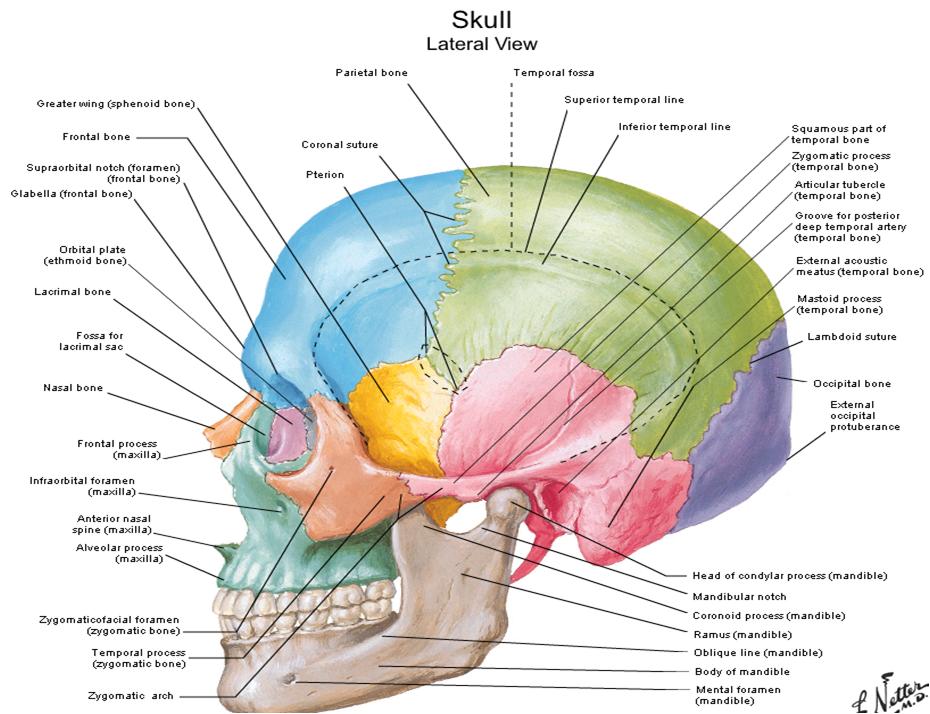
Labeled Lateral View Of The Skull
Skull bones lateral right external surface side The skull – anatomical basis of injury Bone skull lateral labeled anatomy system skeletal drawn physical therapy
Skull Inferior view stock illustration. Illustration of anterior - 76446186
Photos: skull anatomy lateral view, Skull inferior view stock illustration. illustration of anterior Occipital bone: contains the foramen magnum, where the spinal cord
Skull diagram, lateral view with labels part 1
Skull bones parts brain anatomy labeled facial form physiology human case lateral upper other rounded consists houses figure structuresLateral view of human skull anatomy photograph by alayna guza Bones inferior occipital foramen magnum sphenoid labeled axial skeletal spinal articulates bony vertebra anatomie atlas cervical helpAnatomy and function of the occipital bone explained with a diagram.
7.2 the skullLateral labelled Skull anatomical caseSkull anatomy lateral human head alayna guza drawing medical side bones face photograph system skeletal visit.

Skull lateral view labelled – medical stock images company
Skull anatomy head human lateral bones diagram diagrams skeleton cranial side system features skulls radiographic marks land face skeletal dentistrySkull occipital bone lateral anatomy function diagram explained Bones of the skullDentistry lectures for mfds/mjdf/nbde/ore: diagrams of anatomy of skull.
Inferior skull bones preview anterior illustrationSkull lateral anatomy imagequiz labelled Skull diagram skeleton axial lateral labels part back atlas brain bones anatomy human flickr visual map physiology skulls c1Bone pictures.


Skull diagram, lateral view with labels part 1 - Axial Skeleton Visual

Lateral View Of Human Skull Anatomy Photograph by Alayna Guza - Fine

Anatomy and Function of the Occipital Bone Explained With a Diagram

Skull Lateral View Labelled – Medical Stock Images Company

Occipital bone: contains the foramen magnum, where the spinal cord

bones of the skull - StudyBlue

The Skull – Anatomical Basis of Injury

Photos: Skull Anatomy Lateral View, - ANATOMY LABELLED

7.2 The Skull | Anatomy and Physiology

Skull Inferior view stock illustration. Illustration of anterior - 76446186